Glowing plants reveal immune responses around infection sites via two colors · Phys.org · El Autómata
Lunes, 16 de noviembre de 2026
Portada de las 21:20
Todos los periódicos y la radio, en unoEscrito por IA a partir de 26 medios · Cómo se hace
Biología y genética
Phys.org · texto original · Plants & Animals
Glowing plants reveal immune responses around infection sites via two colors
Researchers at the MRC Laboratory of Medical Sciences (LMS) have developed a new way to make glowing biological reporters more accurate, allowing scientists to track gene activity in living plants over days, weeks and potentially even months and years. The approach could help researchers study how plants respond to disease, pests and environmental stress, while opening the door to similar applications in animal and human disease models.
Foto: vía Phys.org
MRC Laboratory of Medical Sciences · Phys.orgPublicado 24 oct, 01:20Ver en phys.org
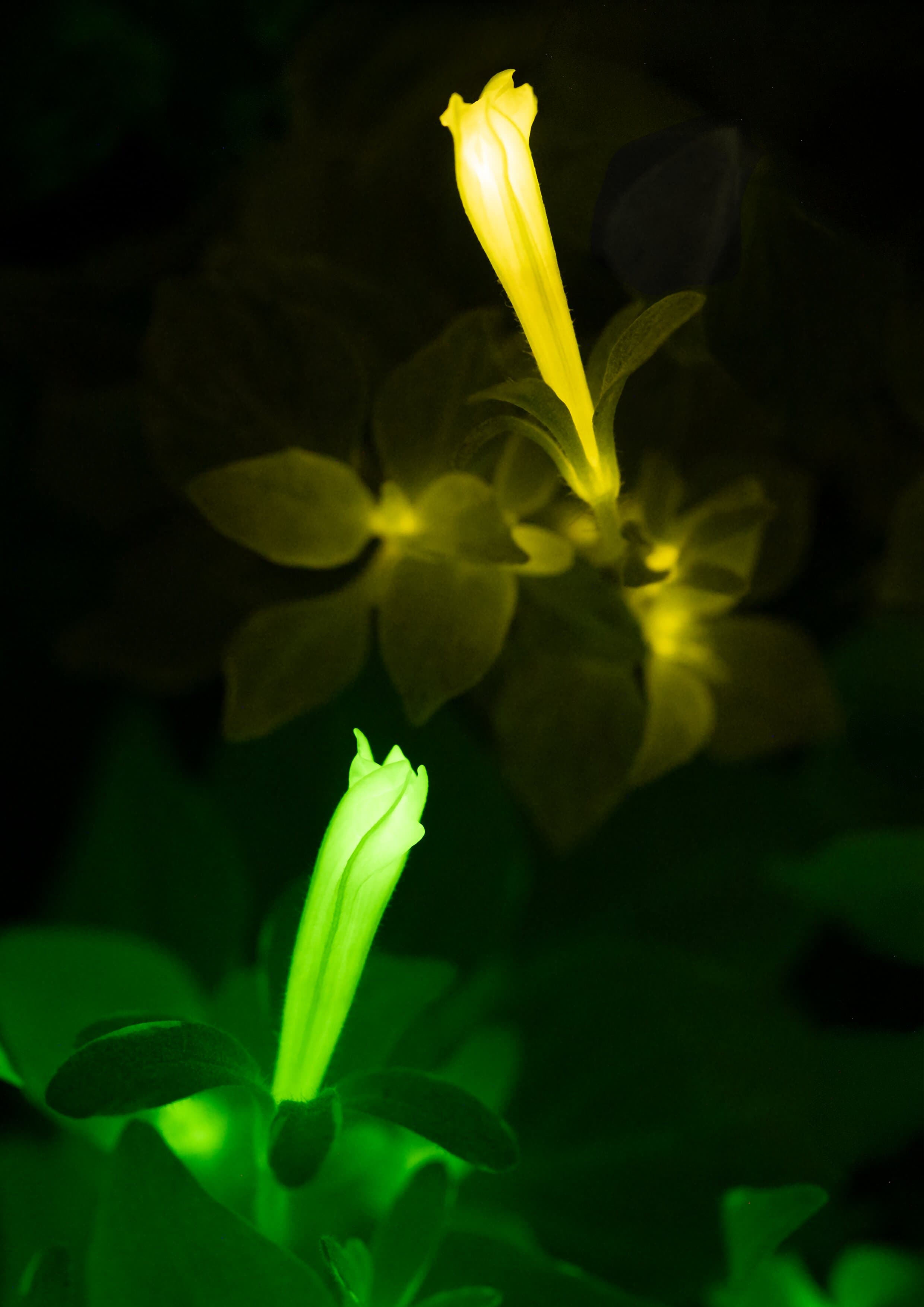
A glowing plant reports its own molecular physiology by a change in its luminescence color. Using change in color instead of change in light intensity as a readout enables more accurate measurement of gene activity by correcting for metabolic effects that influence light emission. Credit: @light.bio
Published today in Nature Plants, the study addresses a longstanding challenge in autoluminescence imaging, a technique that enables organisms to generate their own light without the need for added chemicals or specialized laboratory equipment. The research was led by scientists working in Karen Sarkisyan's group and collaborators from Light Bio and international partner institutions.
Separating biology from background noise
Scientists often use light-producing reporter systems to monitor when genes switch on and off. However, in self-sustaining luminescent systems, light output is affected not only by gene activity but also by changes in metabolism, temperature, oxygen levels and other factors. This makes it difficult to determine whether changes in brightness truly reflect biological activity or simply shifts in the organism's physiological state.
To overcome this problem, the researchers engineered a new red-shifted version of a fungal luciferase and paired it with a green luminescent reporter. By measuring the ratio between the two colors rather than overall brightness, they created a system that remains reliable even when metabolism changes.
The approach effectively transforms autoluminescence from a qualitative indicator into a quantitative measurement tool, providing a more accurate picture of what is happening inside living organisms.
Three-day-long timelapse of 42–44-day-old Arabidopsis thaliana plants. Luz593-expressing plant was used as a control, defense signals ratiometric reporter—pWRKY70 ratiometric reporter. Credit: Nature Plants (2026). DOI: 10.1038/s41477-026-02437-y
Watching plant defenses in real time
The team demonstrated the technique in plants, tracking key defense and immune signaling pathways. Using the new dual-color reporters, they were able to monitor hormone-driven responses over extended periods and detect localized immune activity around sites of infection, even when differences in metabolism caused large variations in overall brightness.
The system worked across both fungal and bacterial bioluminescence pathways, suggesting it could be adapted for a wide range of biological applications.
"One of the biggest limitations of self-sustained luminescence has been that the brightness of the signal depends on both gene expression and metabolism. By introducing a second color as an internal reference, we can separate those effects and obtain a much more reliable readout of what's happening inside a living organism," said Sarkisyan, group head at the LMS and co-corresponding author of the study.
"This turns autoluminescence from a qualitative tool into a quantitative one. In practice, it means researchers can monitor molecular processes over long periods simply by taking photographs in the dark, without expensive equipment or invasive treatments."
From glowing plants to future biomedical applications
The ability to monitor biological processes continuously and noninvasively could support the development of "sentinel" plants that quantitatively report nutrient levels, environmental conditions or disease through changes in color of their light emission. The technology may also help researchers study plant-pathogen interactions and crop health under more realistic field conditions.
While the study focused on plants, the researchers believe the underlying technology has much broader potential. Looking further ahead, the team is exploring ways to adapt similar approaches for use in animal tissues and disease models. This could eventually enable researchers to monitor hormone activity, gene regulation and disease-related processes over extended periods using simple imaging methods.
The work was highlighted by Nature Plants in an accompanying Research Briefing, which described the new ratiometric reporter system as a solution to the variability that has previously limited quantitative applications of fungal bioluminescence. Senior editor Guillaume Tena noted that the new red-shifted luciferase provides an internal control that allows researchers to distinguish genuine biological signals from metabolic fluctuations.
Publication details
Viktor V. Morozov et al., Metabolically robust ratiometric autoluminescent reporters, Nature Plants (2026). DOI: 10.1038/s41477-026-02437-y
Bachelor's in mathematical biology, Master's in creative writing. Well-traveled with unique perspectives on science and language.
Full profile →
Citation:
Glowing plants reveal immune responses around infection sites via two colors (2026, October 23)
retrieved 16 November 2026
from https://phys.org/news/2026-10-reveal-immune-responses-infection-sites.html
This document is subject to copyright. Apart from any fair dealing for the purpose of private study or research, no
part may be reproduced without the written permission. The content is provided for information purposes only.