Long-overlooked Alzheimer’s hypothesis makes a comeback
Decades after it fell out of fashion, researchers return to an idea linking the disease to the loss of crucial neurons
Decades after it fell out of fashion, researchers return to an idea linking the disease to the loss of crucial neurons
A version of this story appeared in Science, Vol 394, Issue 6825.Download PDF
Progress
1.0x
Volume is at 50%
00:00
1.0x
In the mid-1970s, a psychiatry researcher named Raymond Bartus amassed a cadre of aging rhesus monkeys, retired breeders from labs all over the United States, to test a compelling new idea about the origins of Alzheimer’s disease.
A rash of research in the U.S. and United Kingdom implicated the brain’s cholinergic system, made up of neurons that produce a chemical called acetylcholine. Though this neurotransmitter is normally widespread in the brain, autopsy studies found the brains of Alzheimer’s patients mysteriously depleted of it. Researchers also reported severe damage to the nucleus basalis of Meynert, nestled in a deep brain region called the basal forebrain, from which cholinergic neurons shoot projections into areas important for memory and learning.
On the Michigan campus of the drug company Parke-Davis, Bartus tested his monkeys using a complex contraption of his own design, in which the animals could claim a piece of food by correctly recalling which of nine windows they’d seen a light flash in. He found the old monkeys’ short-term memory eroded in ways that closely mirrored early symptoms in Alzheimer’s patients. Their performance improved, though, when he gave them physostigmine, a drug isolated from an African bean that keeps acetylcholine from breaking down. Conversely, young monkeys struggled on the memory test just like older animals when he gave them scopolamine, a drug that blocks cholinergic signaling.
As a drug company scientist, Bartus operated at a distance from the academic neurologists and pathologists leading the human studies. But “nobody [was] talking to each other,” he recalls, so he took it upon himself to cobble together the mounting evidence and top it off with his monkey findings. His 1982 paper in Science became the first to articulate “the cholinergic hypothesis of geriatric memory dysfunction.” It held that loss of cholinergic neurons in the basal forebrain provoked age-related memory loss, and that Alzheimer’s might be conceived of as a more drastic failure of the same system.
No one assumed cholinergic dysfunction was the only thing happening in the brain during Alzheimer’s. It certainly did not explain the signature lesions—sticky “senile” plaques and tangled protein strands—in the brains of those who died with the disease. But the hypothesis offered an intuitive treatment target at a time when investigators had come to suspect that the loss of neurotransmitters could drive neurodegenerative diseases. In the early 1970s levodopa, a medication that stood in for the dopamine lost in Parkinson’s disease, proved revolutionary for patients, relieving tremors and restoring movement. It seemed just a matter of time before similar drugs would emerge to treat Alzheimer’s.
Soon enough they did, but acetylcholine boosters were never the home run levodopa was. The cholinergic hypothesis was barely a decade old when it faced a juggernaut of a rival: the idea that beta amyloid—the sticky protein that makes plaques in the cortex, or brain’s outer layers—was the likely root cause of the disease. Cholinergic research “was already falling out of fashion before the first drugs were even approved,” Bartus laments.
A generation later, the amyloid hypothesis and the treatments it inspired have struggled, whereas evidence supporting a role for cholinergic neurons has strengthened. Now, the field is rediscovering the hypothesis it left behind. “People are returning to other ideas, other cell types, other cellular changes that are affecting cognition,” says neuroscientist Hongkui Zeng of the Allen Institute.
Bartus’s paper was an inspiration to Ezio Giacobini, an Italian pharmacologist working in the U.S. In the 1980s, Giacobini pioneered the development of molecules inhibiting an enzyme that breaks down acetylcholine, keeping more of the essential chemical in the brains of people whose basal forebrain cholinergic neurons were dying. Starting in the 1990s, a series of drugs in this class, known collectively as cholinesterase inhibitors, became the first Alzheimer’s treatments approved by the U.S. Food and Drug Administration (FDA), and three—donepezil, rivastigmine, and galantamine—remain in wide use today. But they don’t work very well or for very long. Even Bartus, a lifelong champion of the drugs, took pains to refer to them as “palliative.”
An experience early in Giacobini’s studies underscored their limitations. He dosed himself with the compound that had revitalized Bartus’s forgetful monkeys: physostigmine, notorious in West Africa for its use as a poison. “Half an hour later, I was vomiting,” Giacobini recalls. “It was too violent, too violent.” Acetylcholine affects many bodily systems, resulting in sweating, salivating, and gastrointestinal distress when levels are boosted. The drugs he developed were gentler, but at doses strong enough to make a difference, the side effects were still formidable.
One perceived weakness of the cholinergic hypothesis was that it could not explain why basal forebrain neurons started to die in the first place. The emerging amyloid hypothesis, on the other hand, suggested a causal chain: In families with inherited forms of Alzheimer’s, genetic mutations favored buildup of sticky amyloid protein in the brain, setting the stage for a complex cascade of damage. Mice engineered to carry these mutations also developed memory problems and plaques, convincing many researchers that if amyloid buildup could be stopped, so could the disease.
“To be fair, if I were in their shoes at that time, I would have probably also said, ‘Holy shit, we’ve solved it,’” says Taylor Schmitz, a neuroscientist at Western University who studies cholinergic function in people and mice.
By the 1990s, the golden age of neurotransmitters was giving way to a fascination with disease proteins—beta amyloid and the tangle-forming protein called tau—and with the areas in the cortex that saw the earliest damage associated with these proteins. Bartus began to feel like an “obligatory invitee” at conferences. He abandoned his cholinergic research in 2000, but not before warning in a paper that although the amyloid findings were indeed thrilling, it might be years or decades before they translated into therapies. In the meantime, the bird in hand—cholinergic drugs—needed to be refined and improved, he argued.
Instead, drugmakers lost interest, shelving agents with novel mechanisms that had shown real promise in trials. Doctors continued to prescribe cholinesterase inhibitors because they were all anyone had.
Some strained attempts to merge the amyloid and cholinergic hypotheses only added to the backlash, says neuroscientist Marsel Mesulam of Northwestern University, who first identified the nucleus basalis of Meynert as the hub of cholinergic neurons. Cholinergic research came to be perceived “as sort of a sideshow” in Alzheimer’s research, he says, “which is unfortunate.”
That is not to say all interest dried up. Pathologists published the first digital maps of the basal forebrain, allowing many more people to study it. Researchers noted a loss of basal forebrain cholinergic neurons in people with dementia due to Parkinson’s and Lewy Body disease, suggesting their failure might be a more general feature of neurodegeneration. (Cholinesterase inhibitors are now used to treat both conditions.) Mesulam discovered abnormal tau inside cholinergic neurons in people who died with cognitive impairment, a hint that disease proteins might have a role in weakening them. And cholinesterase inhibitors appeared to help preserve brain structure and brain blood flow in Alzheimer’s patients, suggesting the drugs might have more than just palliative effects. Mesulam produced periodic review articles revisiting the unfashionable hypothesis and incorporating any intriguing new threads.
Schmitz was a graduate student in the 2010s when he began to read Mesulam’s papers. “Marsel is a very competent scientist, but he also writes very beautifully,” he says. What Schmitz read convinced him that what had happened in the Alzheimer’s field—the abrupt pivoting from the subcortical brain structures to the cortex, from neurotransmitters to amyloid and tau—“was a little tragic.” Lost along the way was the question of where Alzheimer’s begins in the brain. The conventional wisdom now pointed to a region called the entorhinal cortex, where tau is first seen accumulating.
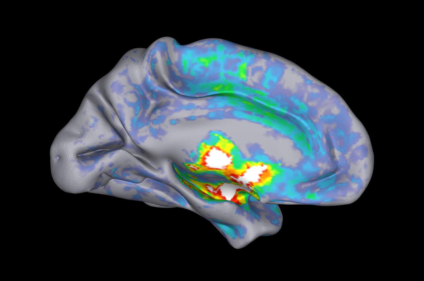
In 2016, Schmitz and neuroscientist Nathan Spreng boldly challenged that with data from the Alzheimer’s Disease Neuroimaging Initiative, a long-term study that uses magnetic resonance and positron emission tomography (PET) imaging to watch the disease process unfold in living people. Degeneration in the basal forebrain predicted later damage in the entorhinal cortex, they reported, not the other way around.
The provocative finding landed just as the first clinical trials were showing no benefit from experimental antiamyloid drugs, designed to clean up plaques in the brain. It seemed some essential element might be missing from the amyloid hypothesis. Other studies were drawing attention back to cholinergic neurons of the basal forebrain: Manipulating these cells with light in transgenic mice revealed how powerfully and precisely they influence perception and learning. New PET tracers let researchers assess real-time cholinergic function in sick and healthy people. And groundbreaking imaging methods finally allowed scientists to see these neurons in all their sprawling glory.
“This is it: the nucleus basalis of Maynert,” says neuropathologist Melissa Murray of the Mayo Clinic Florida, unwrapping a slice of preserved human brain and pointing out an inconspicuous brown dot. “It’s this tiny little nucleus. And yet it has such power because it sends these cholinergic projections all throughout the cortex.”
Pathologists like Murray have known for decades that these specialized neurons are long. But in 2014, when a group at Johns Hopkins University began to use a technique called sparse genetic labeling to visualize individual basal forebrain cholinergic neurons in the mouse brain, even the field’s veterans were startled by the scope of their branching. In the mice, each cell’s axons measure about 50 centimeters if stretched out in a line. A human cell’s axons, laid out the same way, have 100 meters of total projections, the researchers calculated.
Anchored in the basal forebrain of a mouse, a single cholinergic neuron sends bushy projections through the cortex, reaching regions key to emotion, attention, and memory. Cells like this one are among the earliest to die in Alzheimer’s and other neurodegenerative diseases. Scientists think their outsize dimensions and extreme energy needs leave them—and the brain areas they serve—susceptible to age-related damage.
Their staggering dimensions offered clues as to why these cells might be selectively vulnerable in Alzheimer’s and other neurodegenerative diseases. “It’s like the cell bodies are at ground control in Houston, Texas, and their axon terminals are at low-Earth orbit,” Schmitz says. This means they require extraordinary amounts of energy to signal effectively. And they’re naturally more exposed: Compared with other neurons, they have less myelin, a fatty coating that insulates and protects the cells. In June, Schmitz and his colleagues published a sweeping account of the many evolutionary and developmental pressures that, they argue, set these neurons up to fail.
They aren’t the only long-projecting neurons Alzheimer’s researchers worry about. Murray also studies spindly cells that produce the neurotransmitter norepinephrine and originate in a separate deep-brain structure called the locus coeruleus. She has linked specific patterns of Alzheimer’s symptoms to damage to the locus coeruleus and the nucleus basalis of Meynert. The question is not whether these areas and their special neurons are important, she says. It’s “why are they being devastated?”
Schmitz thinks there may not be a universal trigger. Any impairment to the brain’s ability to take up glucose, including age-related vascular changes that reduce blood flow, could hobble these already overtaxed cells and wipe out their branching networks in the cortex. Established Alzheimer’s risk factors such as smoking, sleep disturbances, chronic inflammation, and diabetes could all set the process in motion. Changes to lipid metabolism caused by known Alzheimer’s risk genes like APOE4 could put long-projecting neurons on a path to degeneration as well.
But it’s still not clear why some people can resist these assaults and others progress to dementia. This is where the field needs to focus, Schmitz and Murray both say. “If the brain is endowed with endogenous mechanisms of resilience, how do you drug that?” Schmitz asks. “Rather than just come in and mop up the pathology?”
Giacobini was a schoolboy in Turin, Italy, when the 1936 Nobel Prize in Physiology or Medicine was awarded for pioneering studies of acetylcholine. At 97 he is still publishing, and he has lately had the pleasure of seeing his life’s work on cholinergic drugs vindicated.
Earlier this year at a conference in Copenhagen, Giacobini and University of Southern California psychiatrist Lon Schneider presented an analysis of cognitive outcomes from randomized trials of the main Alzheimer’s drug classes: cholinesterase inhibitors and the newer antiamyloid antibodies. The antibodies are termed “disease-modifying” for their amyloid-lowering effects, whereas cholinergic drugs are deemed “symptomatic” treatments. But their effects were roughly comparable, the researchers found. “We show the same effect [size], only that the cholinesterase inhibitor effect is longer,” Giacobini says. If a drug modifies cognition for years, “isn’t that perhaps disease-modifying?”
Faced with the limitations of antiamyloid drugs, researchers seem to be taking cholinergic agents more seriously. This year “cognition enhancing” therapies, a label that includes most drugs targeting the cholinergic system, were the fastest growing category in the pipeline of Alzheimer’s drugs. Some trials are testing these in combination with other drugs, including an antiamyloid antibody. “Whatever is going to happen in Alzheimer’s therapy,” Mesulam says, “cholinergic drugs will be part of the cocktail.”
Some teams are working on drugs that stimulate specific receptors on cholinergic neurons known to affect cognition, rather than bathe the system in acetylcholine. Medicinal chemist Abraham Fisher, president of AD/PD, an annual Alzheimer’s and Parkinson’s research conference, developed one such agent, NSC001, which is being tried in phase 2 studies to treat people with mild-to-moderate Alzheimer’s. Because the receptors it targets are typically preserved even late in the disease, NSC001 might work in more advanced patients as well, Fisher thinks.
Xanomeline, a drug that activates two types of cholinergic receptors, was tested during the 1990s to improve cognition in people with Alzheimer’s. It seemed to help, but was set aside because of stomach effects. Researchers later combined it with a second drug to block those, and in 2024 the combination won FDA approval for schizophrenia after trials showed it could also treat psychosis. Now, it’s being tested to treat psychiatric and cognitive symptoms in Alzheimer’s patients, with the first phase 3 trial results expected late this year.
Whatever is going to happen in Alzheimer’s therapy, cholinergic drugs will be part of the cocktail.
Other drugs in development target p75, a receptor expressed heavily by basal forebrain cholinergic neurons. P75 helps the neurons form new connections and prune unnecessary ones, but can also prompt their degeneration under stress. Partially blocking this receptor, it’s hoped, can prevent them from dying.
At an Alzheimer’s meeting in July, Stanford University neurologist Frank Longo presented an encouraging analysis from phase 2 studies of a p75-targeting drug. The molecule makes “synapses resilient at a fundamental level,” he says. But Longo and the company he founded don’t emphasize its connection to the cholinergic system.
In many corners of the drug development world, the cholinergic brand remains “tainted,” says Elizabeth Coulson, a neuroscientist at the University of Melbourne who is developing another p75 therapy. Coulson says she’s keen on changing that: “I’ve taken it on as a crusader, almost, to convince everyone that the cholinergic system is important.” Recently, her research has shifted toward how basal forebrain cholinergic signaling drives the glymphatic system, the fluid- and waste-clearance network that becomes disabled in Alzheimer’s, perhaps worsening the disease.
Regardless of whether new drugs succeed, the renaissance in cholinergic research has motivated more scientists to learn what makes cholinergic neurons and other cells like them more vulnerable in the early stages of disease. And it has reoriented a field long obsessed with the cortex—the seat of cognition and ground zero for amyloid buildup—toward less explored deep-brain regions.
That’s a good thing, says Zeng, who has mapped basal forebrain cholinergic neurons in mice, and whose Allen Institute colleagues recently mapped the fabulously long neurons of the mouse locus coeruleus. The institute’s scientists are identifying thousands of cell types in the mouse brain, and roughly 80% originate in subcortical regions. These “are really important also for cognitive and neuromodulatory function,” she says. “It’s not just the cortical neurons.”
Neuroscientist Li-Huei Tsai of the Massachusetts Institute of Technology is a convert to that view. In 2024, while investigating a type of noninvasive brain stimulation to improve glymphatic clearance of disease proteins, Tsai saw the intervention didn’t work when cholinergic signaling in the basal forebrain was deliberately disabled. “This shows that cholinergic signaling is very essential for what we observed,” Tsai says. “When science comes from multiple different directions, but they all converge on the same system, it’s particularly compelling.”
Schmitz has done much to pull the disparate threads of this science together, uniting his fellow imaging experts with the anatomists and the drug developers. This summer he hosted a conference that featured Mesulam and Giacobini as speakers alongside researchers who, like Tsai, stumbled into the field from other directions.
Now 46, Schmitz finds it remarkable how well the hypothesis, which came into the world at about the time he did, is holding up. Mesulam isn’t surprised. “The cholinergic system is not something that is fashionable or not. It’s a reality,” he says. “So why would you not want to research it?”
Texto de Science tal como lo publicó, leído de su página y mostrado aquí con el formato de El Autómata. El original, en science.org · Volver a la noticia de El Autómata